In most cases, a woman who finds herself in a position is interested in the question of which of the two is true: a monthly period or an ultrasound period. And if experienced representatives of the fair sex do not experience problems with determining the age of the fetus, then for the first time pregnant women do not have a clear idea of \u200b\u200bthe differences between obstetric and gestational terms.
When diagnosing pregnancy, the gynecologist announces the obstetric term in weeks. An important feature is that the reporting point is the first day of the menstrual cycle. As you know, conception occurs during ovulation (about 14 days). In this situation, in fact, the woman is not yet pregnant at the moment when menstruation begins. That is why, in most cases, the approximate date of birth (PDD) differs by 2 weeks from the actual or delivered by ultrasound downward.
But it is this method that is optimal and is used in obstetric practice. This is true, because the egg starts its development on the first day of menstruation, and then matures and fertilizes, and if not, then dies. Therefore, the obstetric term can be considered the "age" of the egg. Also, menstrual cycles are individual and for different women can be very different from each other. While the 28-day menstrual cycle is generally considered a reference, actual values \u200b\u200bcan vary greatly.
0dV8PW_Yll8
So, many women may have a cycle longer than 28 days, for example, 35. In this case, ovulation occurs on its 16-17 days. Accordingly, if the cycle is less, for example, 21 days, then the release of the egg from the body of the ovary takes place at 10-11 days. To simplify the work of specialists, it is customary to consider the beginning of pregnancy from the first day of the last menstruation, which is called the obstetric period.
According to the results of ultrasound examination
In the case of determining the maturity of the fetus according to the results of ultrasound examination, a controversial situation may arise:
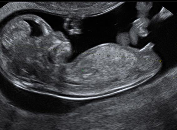
- The temporary course of pregnancy by ultrasound is determined by assessing the development of the fetus, metric indicators, the state of the uterus and the placental barrier (in the II and III trimesters). One of the indicators is the CTE (coccygeal-parietal size), which is almost the same in different fetuses at the initial stages of development. Pay attention to the size of the ovum in the first 12 weeks of gestation. The most accurate is the number of weeks established by ultrasound in the first trimester of pregnancy (up to 12 weeks). After the indicators may vary up or down due to the individual characteristics of the development of the unborn child.
- As a rule, the embryonic date established by ultrasound indicates the period from the moment of fertilization of the egg with male material to the present, therefore, it is considered in fact correct. Most often, there is a discrepancy of about 2 weeks between the PDD by ultrasound or calculated on a monthly basis. But certificates of incapacity for work and other documents are issued based on the obstetric age of the fetus, which is indicated in the mother's passport and documentation in the antenatal clinic.
HAPm0O1ujjw
Conclusion and conclusions
Be that as it may, it is almost impossible to establish exactly when a child was conceived and when he was born. Even knowing the exact date of the "decisive" sexual intercourse, one cannot be sure that fertilization took place on that very day, since sperm cells can exist for 24 hours. Moreover, there are many factors that provoke labor earlier than expected.
If we consider the question of the correctness of the period for menstruation or ultrasound, it should be said that both are true, but traditionally obstetricians are guided by the first option. This avoids confusion in the future and considers possible delivery on the specified dates. But, according to statistics, not every woman gives birth on the day established by specialists. It is considered normal to run at 4 weeks (from 38 to 42 weeks of gestation, respectively).
Pregnancy is one of the most beautiful periods in the life of the fair sex. It is worth noting that medicine knows two options for calculating the time of bearing a fetus in the uterus: obstetric gestational age and real.
How does it all start?
To begin with, it is worth talking about how fertilization occurs. Around the middle of the month, the female egg leaves the follicle and slowly moves along. It is here that it meets the male cell. Further, the chromosomes merge, and conception occurs. Having descended into the uterine muscle, the ovum is introduced into the endometrium, and from that moment it can be considered that the pregnancy has taken place.
Determining the duration of pregnancy
When a woman realizes that she is in an interesting position, her initial task is to determine the date. The gestational age is calculated by week. Usually, the time span during which the baby is in the mother's womb is 40 weeks. A slight shift in one direction or another is considered normal and does not require any correction. Doctors distinguish between obstetric and real pregnancy.

Real time of carrying a baby
This period is counted from the moment when ovulation occurred. The release of the egg from the follicle is the day from which the actual gestational age is calculated. Most women's clinics that monitor the course of pregnancy use this particular calculation method. If you decide to take a blood test to determine the content in it, then you will also be provided with a result that indicates the real value of the period.
Obstetric gestational age
This time period begins its countdown from the first day of the last spotting from the genital tract of a woman. To calculate the estimated date of birth of the baby, this is the term used. Also, many of the fair sex use this method in order to establish the period of pregnancy. That is why so often women have discrepancies with the calculation made by the doctor.
Obstetric gestational age and real
In most cases, the difference between these counting methods is two weeks. With a standard female cycle of twenty-eight days, the release of the egg from the ovary occurs exactly two weeks after the start of the last menstruation.
However, not all women have a standard cycle length. For example, some women ovulate one week after the start of their last period. In such cases, the difference between obstetric and real terms will be one week.
If a woman's egg release occurred three weeks after the start of the last menstrual period, then in this case the obstetric gestational age and the real one will have a difference of twenty-one days.
All situations described are normal. That is why the duration of pregnancy by week should be set taking into account the length of the woman's menstrual cycle. It is impossible to equate all the fair sex according to generally accepted standards. This can lead to an incorrect calculation of the period of bearing the baby.
pregnancy by ultrasound

There are situations when a woman cannot name the date of the first day of her last period. This situation often occurs if a woman has recently given birth or is breastfeeding. In such cases, the fair sex is recommended to undergo diagnostics with an ultrasound machine (ultrasound).
A short period of pregnancy, which is not yet possible to establish with a manual examination, is easily diagnosed by ultrasound. It is worth noting that a specialist can determine the presence of a woman in the uterus starting from the fourth obstetric week. All measurements and definitions of the term are calculated in this obstetric way.

Instead of a conclusion
If you are unsure of how to calculate your pregnancy, ask your doctor. In most cases, it is enough to know the date of the last period and the length of the woman's menstrual cycle. If necessary, an ultrasound examination is also prescribed. Calculate using the same method as a specialist. Only in this case you will not encounter discrepancies and will not get into a controversial situation.
The availability of ultrasound diagnostics allows a woman who has just learned about the onset of pregnancy not to speculate about the term, but to find out exactly how her baby is developing. Naturally, when visiting an ultrasound office, a woman roughly suggests how long the doctor will call, but it happens that her expectations are not met.
How often does this happen, how can you explain the situation when the ultrasound period differs from the monthly period, or the size of the fetus differs from the standard for a specific period?
How much can you trust ultrasound?
Ladies who really want to get pregnant want to know about the accomplishment of a miracle in their lives as early as possible, literally in the first days of the delay. Of course, you can always buy a pregnancy test. But even supersensitive tests are only able to accurately give a specific answer to the question "has a pregnancy occurred?" They are able to answer yes or no, but more precise methods will be required to set a timeline.
Positive pregnancy test
During a manual examination of a pregnant woman, an obstetrician-gynecologist may notice that the uterus is loose and slightly enlarged. But this phenomenon is observed before menstruation. To accurately clarify the situation, you need an ultrasound scan.
Ultrasound is able to accurately determine pregnancy when the level of hCG (chorionic gonadotropin) in a woman's blood exceeds a thousand units. In this case, the doctor is already able to see the ovum in the uterus (with multiple pregnancies - two or three ovules). The longer the period, the more complete the study shows - the doctor ascertains the presence of not only the ovum, but also the yolk sac in it, or even sees the embryo and its heartbeat, and will be able to measure the CTE of the embryo (distance from the tailbone to the crown).
If your menstrual cycle is more than 30 days, the ultrasound and menstrual periods may differ significantly.
Often there is such a situation: a woman comes for an ultrasound scan, the doctor is interested in the date of the last menstruation, conducts a study, and then announces that the pregnancy, most likely, has definitely been frozen - the embryo and its heartbeat are not visualized, the fetal egg is visible, the size of which is smaller than it should to be. The doctor may be wrong if the woman has had late ovulation. In a significant part of women, the menstrual cycle is not the standard 28 days, but 33, or even 35-40. This means that conception did not occur on the 14-16th day of the cycle, but a week or two later, and the woman simply came to the ultrasound scan too early, so the fetus was not seen. Most likely, the pregnancy will not be frozen and the fetus is developing normally, just the study should be repeated in a couple of weeks, so as not to be mistaken for sure. It is also possible to explain the situation when pregnancy is not visible at all on ultrasound: it is likely that the level of hCG in the blood is less than that at which pregnancy can be seen.
Differences in terms of pregnancy by menstruation and by ultrasound

Obstetric gestational age is calculated by week
All obstetricians-gynecologists use the term "obstetric term" when determining the gestational age. This allows all gynecologists in the world to speak the same language. It is conducted in weeks, the countdown of which starts from the first day of the last menstruation. The first and second obstetric weeks are those weeks during which an egg is still maturing in the body, which will subsequently leave the ovary during ovulation and meet with the sperm.
Thus, a woman who has a standard menstrual cycle of 28 days and comes to the doctor with a delay of one week will be diagnosed as pregnant at exactly five obstetric weeks. At the same time, with an ultrasound examination, a slightly shorter period can be set - at three or four weeks. How justified is this and why does the term not coincide? This situation is normal, since the ultrasound does not aim to determine the exact period in weeks, but to determine how many weeks from conception the fetus is developed in its parameters (its gestational age). It is important to understand the difference between the two. Therefore, a discrepancy of 1-2 weeks between the period for menstruation and for ultrasound in favor of menstruation (i.e., the period for menstruation is longer, and for ultrasound less) is a common occurrence, there is nothing wrong with it.
How to determine the duration of pregnancy with an irregular cycle?
Some women suffer from hormonal disorders and, as a result, an irregular cycle. With pathologies such as polycystic ovaries, multifollicular ovaries, an excess amount of male sex hormones, menstruation may not come for several months. Nevertheless, ovulation with such violations still occurs from time to time, which means that the chances of getting pregnant in such women are almost the same as in the rest.

The girl recalls the date of the last menstruation
If pregnancy is undesirable for you, the use of contraception is mandatory, even if you suffer from an irregular cycle and are absent for several months.
Often, women with irregular cycles do not consider it necessary to use protection, believing that they will definitely not be able to get pregnant. In this case, you can find pregnancy in the third or even fourth month, because the absence of menstruation for several months becomes a kind of norm. The correct calculation of the obstetric term in such a situation is practically impossible, therefore, when determining the term, the doctor can rely solely on ultrasound data. If the fetus is not too large or too small, an ultrasound scan can determine the gestational age of the fetus within 2-3 weeks. The date of birth will also be established by an unconventional method - counting 40 weeks from the first day of the last menstruation, and based on ultrasound data.
In what cases is the term for ultrasound ahead of obstetric?
Currently, all pregnant women in Russia are entitled to undergo a free ultrasound examination three times. Usually they are prescribed between 11 and 14 weeks, between 18 and 22, and between 32 and 34. Each of these ultrasounds has its own purposes (the first is to determine whether they have seen gross malformations of the fetus and genetic abnormalities, the second is to control the development of internal organs, the third is to determine the state of the placenta, the position of the fetus and its approximate weight). However, with each examination, the doctor determines how many weeks the fetus has developed. And often this period is ahead of the obstetric one. For example, a woman knows for sure that her pregnancy is 20 weeks, and the doctor indicated in the conclusion that the size of the fetus corresponds to 22 weeks. Why is this situation developing? There is no mistake in it. This can be explained as follows:
- The fruit is large. The large size of the fetus, ahead of its gestational age, is not a pathology. Just like all people, the fetus already in the womb has its own individual characteristics.
- The size of the fruit is slightly smaller than it should be. This may well be a variant of the norm, perhaps the baby is just small, especially if his parents are not distinguished by their high growth and impressive weight.
- Obstetric weeks are incorrectly defined. There are cases when the bleeding, which a woman takes for the next menstruation, is in fact a threat of miscarriage when pregnancy has already occurred in the previous cycle. In the people, this phenomenon is called "washing the fetus." It turns out that a woman tells the doctor one date of the last menstruation, believing that conception occurred in this cycle, while in fact it happened in the previous one, and, accordingly, the obstetric period will be longer.
How often is ultrasound wrong?
There is always a possibility of error, both upward and downward, when determining the term for ultrasound. Why is this happening and how many factors are to blame? There are several of them: outdated equipment, insufficient qualifications of a functional diagnostics doctor, confusion with the date of the last menstruation, they did not immediately see the fetus, the individual characteristics of the fetus (too large or too small). The risk of errors can be minimized. It is enough to visit proven medical centers, in which the level of qualifications of doctors and the quality of equipment is beyond doubt, as well as closely monitor your cycle and know exactly the date of your last menstruation.
Dear Olya!
Your assumption that the ultrasound scan determines the obstetric gestational age is not entirely correct. This study is aimed rather not at setting the date, but at determining whether the size of the fetus corresponds to the expected true gestational age.
Calculation of the duration of pregnancy by ultrasound
During an ultrasound examination in the early stages, fetal development indicators are assessed using tables calculated for the embryonic period, which is 2 weeks less than obstetric. There are several such tables, compiled by different authors, which contain data on the average size of the fetus at different stages of pregnancy and suggest an error interval that can reach 2 weeks. The ultrasound machines have built-in programs in which averaged indicators are taken from different tables. It is possible to judge whether the fetus has a developmental delay only if the woman knows exactly when conception occurred, otherwise the specialist can only make a preliminary conclusion, which must be confirmed by additional research.
In the early stages of pregnancy (up to 12 weeks), specialists pay attention to the length of the embryo from the coccyx to the crown (CTE) and the average inner diameter of the ovum. At later stages of pregnancy, the development of the fetus occurs in leaps and bounds, so the tables that the uzist doctor is guided by are designed for the obstetric period, that is, from the first day of the last menstruation.
It is for this reason that discrepancies often arise between the period that was set for the ultrasound scan and the period that passes in all the documentation drawn up in the antenatal clinic. Therefore, very often, uzist doctors give out a certain average result, taking into account the data on the date of the last menstruation, the date of conception, the results of previous ultrasound and data on the development and growth of the fetus, which in most cases coincides with the true gestational age, if it proceeds normally.
How to determine the date of conception?
To determine the date of conception, you need to calculate the time at which you ovulated and analyze which of those days when you had sexual intercourse was closest to the period of ovulation. It is worth noting that it is almost impossible to determine exactly when a child was conceived, but, nevertheless, an approximate calculation can be made.
To determine when you ovulate, you need to know the length of your menstrual cycle. If it is stable and is 28 days, then you will most likely ovulate on day 14 m.ts. If its duration is 29 days, then on day 15 m.ts. With a 30-day cycle on day 16, etc. The data of the first ultrasound scan and the level of hCG analysis from 7.04 indicate that you could have ovulated in the period from 6 to 8 March 2015. If we adhere to these data, then according to the results of subsequent ultrasound, the development of the child approximately corresponds to the embryonic period of pregnancy with a slight excess. But, as mentioned earlier, the development of the fetus has an abrupt nature, therefore, an error of +/- 2 weeks is considered a variant of the norm.
The diagnosis of fetal growth retardation cannot be made on the basis of the difference in gestational age alone. The delayed development of the fetus should be confirmed by the data of regular measurements of the height of the fundus of the uterus and the circumference of the abdomen, which are made at each visit to the obstetrician-gynecologist; study of the level of placental hormones; CTG data (cardiotocography of fetal cardiac activity); and ultrasound in the case of delayed development reveals a low birth weight, disproportionate growth of organs, impaired blood circulation through the umbilical cord vessels and arteries, and other possible deviations from the norm.
Regards, Ksenia
The duration of pregnancy is a significant indicator. It is used to assess how the fetus develops, to find out the estimated date of birth. There are many ways in which a pregnant woman can set a due date (for example, by the date of the last menstruation that began, by ovulation).
Ultrasound diagnostics (USL) deserves special attention. She is appointed during the period of gestation for several reasons. First of all, ultrasound is necessary to confirm the development of uterine pregnancy. Reasons for performing a scan include determining the length of the gestation period.
Features of setting a time limit
With the help of ultrasound diagnostics, the duration of the gestation period can be determined as accurately as possible in the first trimester. In the next trimesters, the information received is not entirely correct. Errors arise due to the constitutional characteristics of the development of the fetus, as well as due to complications that are present and progressing in some women during pregnancy.
How is the duration of gestation determined using ultrasound?
In the first 3 months, when it is impossible to see the embryo, the specialists will recognize the ultrasound time by the calculated SVD of the ovum - the average inner diameter. This parameter is determined by the following algorithm:
- the measurement of the anteroposterior and longitudinal dimensions of the ovum is carried out during longitudinal scanning;
- the width is measured during transverse scanning;
- the arithmetic mean is calculated from the numbers obtained.
At 5.5 weeks. the average inner diameter is characterized by values \u200b\u200bfrom 0.6 to 0.7 cm.Every day the embryo grows with a normally developing pregnancy:
- at 6 weeks the indicator in question already becomes equal to 1.1 cm;
- at 6.5 weeks - 1.4 cm;
- at 7 weeks - 1.9 cm;
- at 7.5 weeks. - 2.3 cm;
- at 8 weeks - 2.7 cm.
When the embryo begins to be visualized, the indicator that allows you to know the length of the gestation period becomes CTE - a size called the coccygeal-parietal.
Determination of CTE with ultrasound
It is determined by a sagittal scan. This parameter means the maximum distance from the coccyx to the outer contour of the head end:
- at 1 month and 3 weeks. CTE is 0.81 cm;
- at 2 months. - 1.48 cm;
- at 2 months. and 1 week. - 2.24 cm;
- at 2 months. and 2 weeks. - 3.12 cm;
- at 2 months. and 3 weeks. - 4.21 cm;
- at 3 months. - 5.11 cm;
- at 3 months. and 1 week. - 6.32 cm;
- at 3 months. and 2 weeks. - 7.67 cm.
In the second and next trimesters, the duration of pregnancy is determined by various fetometric parameters. 
Specialists can take into account the size of the fetal head in circumference, biparietal size, average diameters of the abdomen and chest, size of the abdomen around the circumference, femur along the length.
How long does the ultrasound scan show: obstetric or from the moment of conception?
Obstetricians-gynecologists use in their work such terms as obstetric and gestational (embryonic) stages of pregnancy. There is a slight difference between these concepts. The obstetric period is the number of weeks that have passed since the beginning of the last period. The gestational (embryonic) period is the period that began from the moment of fertilization of the egg.
The period determined by ultrasound is considered embryonic. In obstetric practice, the first concept is widely used. That is why, in order to avoid confusion, experts translate the gestational period into obstetric, adding 2 weeks to it.
If the period calculated according to the ultrasound scan data exceeds the obstetric ...
Theoretically, the gestational period is a couple of weeks less than obstetric. However, sometimes ultrasound diagnostics shows something completely different. Some women note that their ultrasound pregnancy period is longer than obstetric... This is a perfectly acceptable phenomenon.
The difference is explained by a decrease in the accuracy of determining the timing as the fetus develops. The most accurate information is provided by an ultrasound scan carried out in the first 3 months of pregnancy. During this period, all women develop a fetus almost the same, therefore, errors in determining the term are minimal. 
In the second trimester, the gestational age can be quite accurately determined by fetometric parameters, but in the third trimester, errors already occur due to the fact that each fetus begins to develop individually, genetic factors are influenced. Errors in some cases are ± 3-4 weeks. In the last trimester of pregnancy, fetometry is recommended not to be used to clarify the duration of gestation, but to determine whether the size of the fetus corresponds to an already known period.
Why is the term specified with the help of ultrasound scan?
Post-term pregnancy is one of the problems faced by women in position. In this condition, the embryonic and obstetric period is longer than the established values. A normal pregnancy lasts 38 embryonic or 40 obstetric weeks. Postterm pregnancy is referred to as factors that increase the likelihood of complications during delivery and lead to an increase in perinatal morbidity and mortality. 
To prevent the consequences of post-term pregnancy, there are certain preventive measures. One of them is the exact establishment of the gestational age based on the results of ultrasound diagnostics (it is desirable that women who are in position should be scanned no later than 20 weeks). Determining the number of weeks also avoids unnecessary stimulation of labor.
Knowing the duration of the gestation period enables the doctor to determine whether the fetus is developing according to the norm, whether there are any deviations. Another reason why you need to know the exact number of weeks is the need for a woman to undergo screenings, to take various tests at a certain time (if you take one or another test later or earlier, you can get an unreliable result).
In conclusion, it should be noted that ultrasound scanning is a fairly simple way to determine the length of pregnancy. The method provides the most accurate information in the first trimester. It is from the period calculated at the beginning of pregnancy that doctors start off in the future. It is also worth noting that many mothers are interested in the safety of ultrasound. Ultrasonic waves can be harmful. However, modern devices have a minimal effect on the body, so the diagnostic method is considered safe for both the expectant mother and the fetus.